
3D CBCT Imaging for Endodontics in Oakton, VA
At Endodontic Studio | Oakton, 3D CBCT imaging is available when additional information is needed to diagnose or plan treatment for complex endodontic conditions.
Dr. Abasin Safi uses limited-field 3D imaging when clinically appropriate to evaluate the tooth and surrounding structures while keeping the area of imaging focused on the region of interest.
CBCT may be particularly useful when evaluating previously treated teeth, root resorption, suspected cracked teeth, complex anatomy, persistent infection, or planning endodontic microsurgery.
Our goal is not to obtain more imaging than necessary, but to use the appropriate technology when it can meaningfully improve diagnosis and treatment planning.
-
Cone beam computed tomography, or CBCT, is a type of dental imaging that produces a three-dimensional view of the teeth, roots, surrounding bone, and nearby anatomical structures.
Unlike a traditional dental X-ray, which provides a two-dimensional image, CBCT allows the area to be viewed from multiple angles and in thin cross-sectional slices. This additional detail can be especially helpful when evaluating complex endodontic conditions.
CBCT is used selectively when the additional information is expected to improve diagnosis or treatment planning.
-
Traditional dental X-rays provide valuable information and are sufficient for many endodontic evaluations. In certain situations, however, three-dimensional imaging can reveal details that may be difficult to appreciate on a two-dimensional image.
CBCT may be helpful when evaluating:
Complex or unusual root canal anatomy
Teeth with previous root canal treatment
Persistent symptoms when conventional imaging does not clearly identify the cause
Dental trauma
Persistent infection or delayed healing
Treatment planning for endodontic microsurgery
The decision to obtain a CBCT scan is based on the individual tooth, symptoms, clinical examination, and information already available from conventional imaging.
-
CBCT imaging allows the tooth and surrounding structures to be evaluated in three dimensions, which can provide important information for diagnosis and treatment planning.
Complex Root Canal Anatomy
Some teeth have additional canals, unusual root shapes, or anatomy that is difficult to visualize on traditional X-rays. CBCT may help identify these variations before or during treatment.
Previous Root Canal Treatment
For a previously treated tooth, CBCT may help evaluate the quality and extent of the existing treatment, identify untreated anatomy, and determine the location of persistent changes around the roots.
This information can be useful when determining whether root canal retreatment, surgery, or another treatment option should be considered.
Root Resorption
CBCT can be especially valuable when evaluating root resorption because it can help determine the location, size, and extent of the resorptive defect and its relationship to the root canal system.
Dental Trauma
Following dental trauma, three-dimensional imaging may provide additional information about the roots, surrounding bone, and supporting structures when conventional images do not fully explain the clinical findings.
Cracks and Fractures
Cracks and fractures can be difficult to diagnose, and many are too small to be directly visible on CBCT.
In selected cases, however, CBCT may reveal changes in the surrounding bone or other findings that support the diagnosis or help rule out alternative causes of symptoms. A cracked tooth evaluation therefore relies on clinical testing in addition to imaging.
Persistent Infection
CBCT can help identify areas of bone loss or inflammation around a root that may be difficult to visualize on a traditional dental X-ray. This can be useful when investigating persistent symptoms or evaluating healing after previous treatment.
Surgical Treatment Planning
Before endodontic microsurgery, CBCT may be used to evaluate the location of the root, the extent of the affected area, the thickness of the surrounding bone, and the relationship to nearby anatomical structures.
This information can help make surgical treatment more precise and predictable.
-
A dental CBCT scan is quick and noninvasive. You will be positioned in the imaging unit while the scanner rotates around your head and captures a series of images.
The scan itself typically takes less than a minute, and you will not feel anything while the images are being taken.
The resulting three-dimensional dataset can then be reviewed in multiple views and cross-sections to evaluate the tooth and surrounding structures in greater detail.
-
Some tenderness or sensitivity to biting is common for several days after root canal treatment. Until the tooth has been permanently restored, avoid chewing hard or sticky foods on that side whenever possible.
In many cases, the tooth will need a final restoration—often a crown—to protect it from fracture and restore normal function. We will communicate with your general dentist regarding the appropriate next step and timing for your permanent restoration.
Follow-up may also be recommended to evaluate healing around the tooth. If a previously treated tooth develops new symptoms in the future, root canal retreatment may sometimes be considered.
-
Endodontists are dental specialists who focus on diagnosing tooth pain and treating conditions involving the dental pulp and tissues surrounding the roots of teeth.
After dental school, endodontists complete additional specialty training dedicated to root canal treatment, complex diagnosis, retreatment, dental trauma, and endodontic surgery. Their practices are limited to endodontic care, allowing them to perform these procedures routinely using specialized technology, magnification, and techniques designed for complex root canal anatomy.
At Endodontic Studio | Oakton, our focus is on accurate diagnosis, thoughtful treatment, and preserving your natural tooth whenever possible.
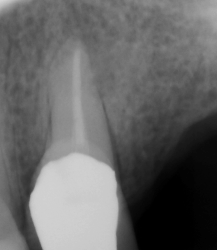
Figure 1
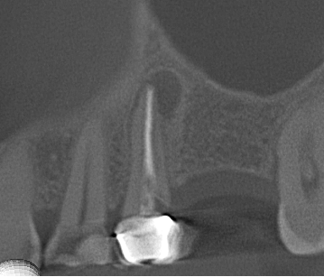
Figure 2
Figure 1 and 2 show the same tooth, but Figure 1 is a 2D image and Figure 2 is showing one view of a 3D image. You can very clearly see signs of infection at the tip of the root when looking at Figure 2 as compared to Figure 1.
Reviewed by Abasin Safi, DMD
Endodontist, Endodontic Studio | Oakton